⚡ In short
A sea star that loses an arm can regrow it as long as part of its central disc remains. Dedifferentiated cells act like stem cells, rebuilding a new arm over months to a year. This article explains the mechanism, the conditions required, and why a species with such regenerative power can still face extinction.
Spot a sea star with one short arm in a tide pool and most people would not give it a second thought. But at the base of that missing arm, a small bump appears within weeks and, over months to a year, grows back into a full arm. Sea stars (echinoderms of the class Asteroidea) are among the few animals that can regenerate a lost arm, and in some species a single arm can even regrow into a whole new individual.
This regenerative ability is more than a curiosity. In a sea star's body, wound cells undergo a kind of "reset" called dedifferentiation, behaving like stem cells - a process studied worldwide, alongside newt limb regeneration and human wound healing, as a model for understanding how animals regenerate.
Yet a mass die-off that swept the North American west coast starting in 2013 pushed some of these regeneration specialists to the brink of extinction. The episode makes clear that an individual's regenerative power and a species' chance of survival are two separate questions. This article draws on primary sources to lay out how sea star arms regenerate, what conditions are required, the latest research findings, and the state of conservation efforts on the ground.
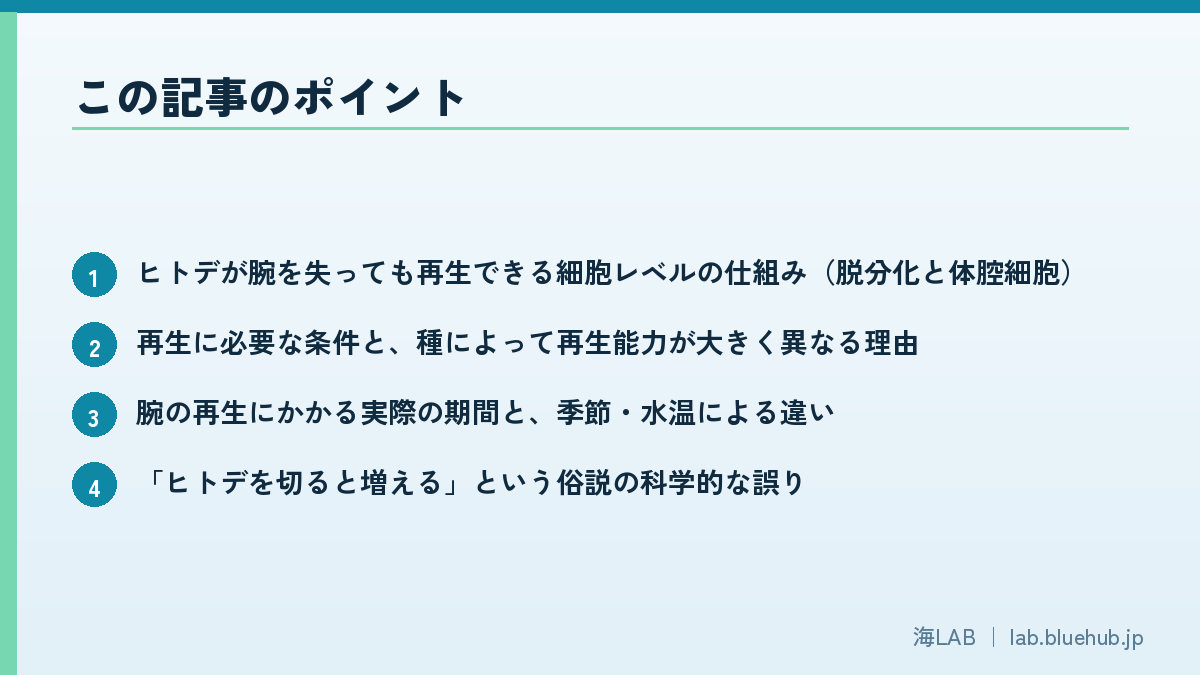
What you'll learn in this article
- The cellular mechanism that lets sea stars regrow lost arms (dedifferentiation and coelomocytes)
- The conditions needed for regeneration, and why regenerative ability varies so much by species
- How long arm regeneration actually takes, and how season and water temperature affect it
- Why the popular belief that "cutting a sea star makes more sea stars" is scientifically wrong
- The case of the sunflower sea star, which went endangered despite its regenerative power, and why
- What sea star regeneration research could offer regenerative medicine
What kind of animal is a sea star?
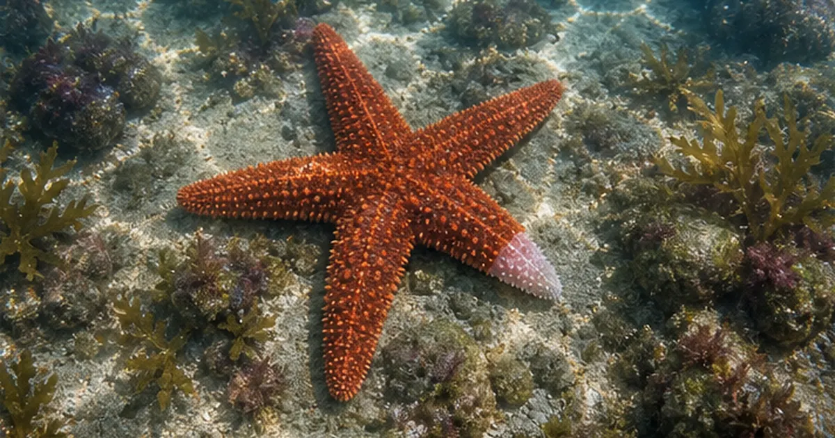
Sea stars are not fish; they are invertebrates belonging to the echinoderms, the same group as sea urchins, sea cucumbers, and brittle stars. Their bodies show five-fold radial symmetry, with arms - usually five - radiating from a central body. They have no backbone or brain-like central organ; instead, light-sensing eyespots sit at each arm tip, and countless tiny tube feet on the underside of the arms handle movement, feeding, and respiration.
Where sea stars fit among the echinoderms
The phylum Echinodermata includes sea stars (class Asteroidea), brittle stars, sea urchins, sea cucumbers, and sea lilies. All share calcium-carbonate skeletal plates near the body surface and a fluid-filled "water vascular system" that drives the tube feet. Sea urchins, which graze on seaweed and contribute to kelp forest loss, are echinoderms too, and some urchin species share sea stars' regenerative ability.
Brittle stars look similar and share the word "star" in their common name, but they are not asteroids - they belong to a separate class, Ophiuroidea. Their arms are thin and long, clearly set off from the central disc, and readily self-amputate (autotomize), regenerating afterward with an ability that rivals sea stars. Research from Hokkaido University found that five-armed and six-armed brittle stars "bulge" differently as they move, showing that something as simple as arm count can shape the coordination of movement.
The diversity of sea stars worldwide and in Japan
According to the US National Oceanic and Atmospheric Administration (NOAA), about 2,000 sea star species live in the world's oceans, adapted to everything from shallow tropical water to the deep sea floor. The waters around Japan, enriched where the Kuroshio and Oyashio currents meet, boast some of the richest marine biodiversity on Earth, and many species are recorded here, from the tide-pool regulars Patiria pectinifera and Linckia laevigata to deep-sea forms.
How a brainless animal controls its body
A sea star has no single central organ equivalent to a brain or heart. Instead, a "nerve ring" circling the mouth and "radial nerve cords" extending into each of the five arms form the skeleton of its nervous system, controlling movement in a decentralized way based on input from light-sensing eyespots at the arm tips and chemoreceptors scattered across the body surface. The water pressure that drives the tube feet comes from the "water vascular system," a pump-like structure that draws in seawater; besides locomotion, this system also handles part of the animal's respiration and gas exchange.
This lack of a single central hub is one major reason a sea star can keep functioning even after losing an arm. In mammals, damage to the brain or heart can halt the whole organism's vital functions, but because a sea star's nervous system and key functions are distributed across its body, local damage rarely proves fatal to the whole animal - laying the groundwork for the arm-by-arm regeneration discussed in the next section.
Common sea stars found in Japan's tide pools
- Patiria pectinifera: a star with short, stubby arms, one of the most commonly seen species in tidal flats and rocky tide pools
- Linckia laevigata: known for its striking blue color, common in the shallows of coral reef areas
- Asterias amurensis: a large species with thick, short arms living on sand and rock
- Astropecten scoparius: identified by its slender, spined arms; it burrows into the sand of tidal flats
How an arm regrows: the cellular mechanism of regeneration
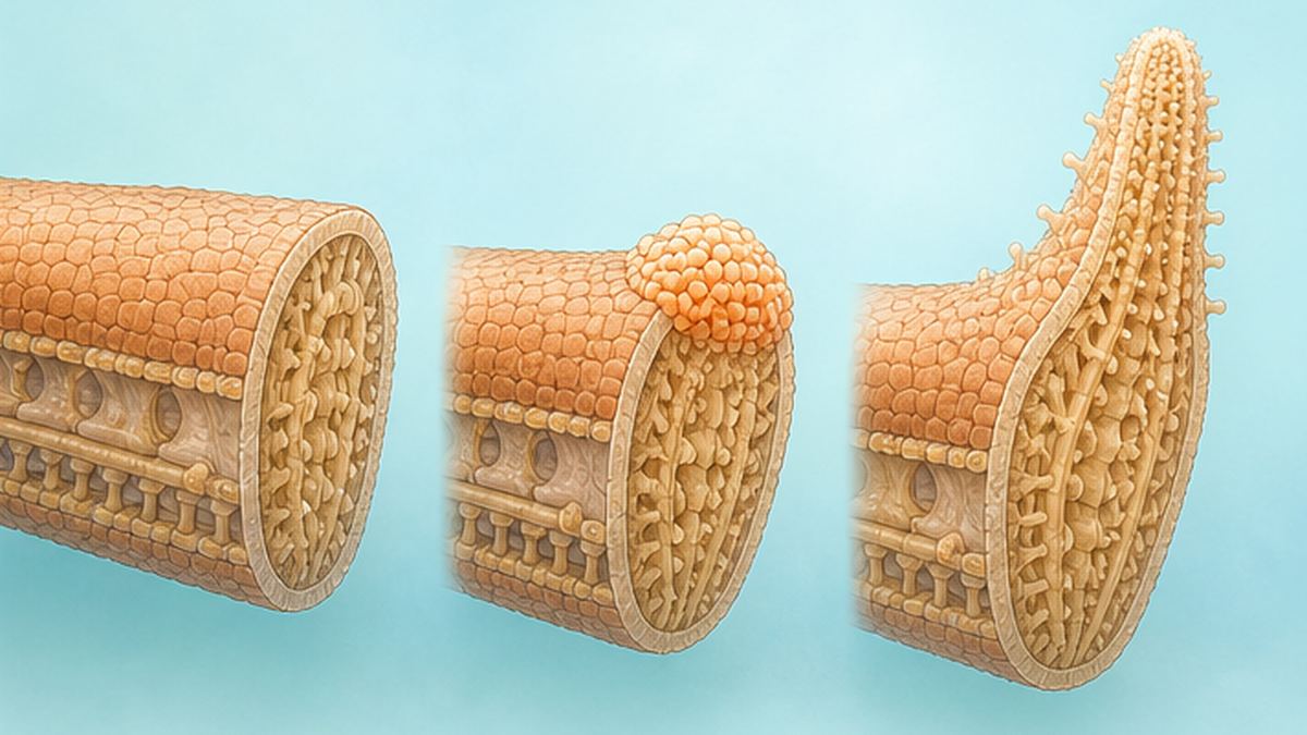
Sea star regeneration proceeds in stages: cells in the damaged tissue first lose their "specialization" and revert to an undifferentiated, stem-cell-like state, then are rebuilt into whatever cell types are needed. This process, called dedifferentiation, is a different strategy from the human approach of mobilizing stem cells held in reserve somewhere in the body.
The role of coelomocytes
A sea star's body fluid contains free-floating cells called coelomocytes, which swarm to a wound to form a clot-like mass and act as immune cells, engulfing bacteria and debris. A 2023 study (Ben Khadra et al.) that closely tracked nerve-cord regeneration reported that a new type of coelomocyte population appears in the later stage of regeneration and supports functional recovery.
Skin regeneration and muscle dedifferentiation
In mammalian wound healing, epidermal stem cells divide actively to close a wound, but sea star skin regeneration works somewhat differently: cells at the wound margin are known to stretch inward to cover the wound. At the same time, muscle cells inside the arm dedifferentiate into mesenchymal-like cells and migrate toward the regenerating tip, a process known as epithelial-mesenchymal transition.
What is dedifferentiation?
- Cells that once had a specific role (muscle, skin, etc.) lose that identity and revert to an undifferentiated state
- These reverted cells then proliferate and differentiate like stem cells, becoming the raw material for rebuilding lost tissue
- Similar dedifferentiation occurs in newt limb regeneration, giving this field common ground with vertebrate regeneration research
Immune response and regeneration as one process
Immediately after a sea star is wounded, coelomocytes rush to the site, forming a clot-like aggregate that both stops fluid loss and engulfs bacteria and other invaders. Animal studies show that if this early immune response fails, subsequent tissue formation stalls - regeneration must first pass through a "keep the wound clean" phase before the "build new tissue" phase can begin. Some researchers compare this to the inflammatory, proliferative, and maturation phases of human wound healing.
Once coelomocytes have covered the wound and fluid loss has stopped, re-epithelialization begins along with formation of a regeneration bud, or blastema. The blastema is a mass of dedifferentiated cells whose fate is not yet decided - a structure also seen in newt limb regeneration and planarian whole-body regeneration. The sea star's blastema receives chemical signals from surrounding tissue and gradually differentiates into specific tissues: muscle, skeletal plates, tube feet, and gonads.
Skeletal plates and pigment come back last
The calcium-carbonate skeletal plates (ossicles) that support a sea star's body form a mesh-like network just under the skin, giving it both rigidity and flexibility. During regeneration, soft tissues - skin, muscle, tube feet - form first, while plate deposition and skin pigmentation lag behind; this is why a newly regenerated arm often looks paler and slightly translucent compared with the rest of the body. Over time, as the plates thicken and pigment cells increase, the arm gradually becomes indistinguishable from the original.
What's required for an arm to regenerate
Most sea star species can regrow one or two lost arms without any trouble. But whether a single detached arm can regenerate into a whole new individual depends heavily on whether a piece of the central disc came away with it. Arm count itself varies by species - five is most common, but multi-armed species such as some Solasteridae have ten or more, and species with more arms tend to be less affected by the loss of one or two.
The disc is the command center of regeneration
A sea star's key organs - part of the digestive glands, the gonads, and the nerve ring that anchors the nervous system - are concentrated in the disc. If an arm is severed with no disc tissue attached at all, in most species that arm cannot build a new individual and eventually dies. Conversely, if even a small piece of disc remains attached, some species can use it as a starting point to regrow the missing arm and eventually develop into a complete individual.
This is closely tied to the fact that a sea star's nervous system is not concentrated at a single point but forms a ring - the nerve ring. If even part of that ring survives, it may be able to rebuild the nervous system's function from there. Conversely, if the nerve ring is entirely lost, no amount of coelomocyte activity or muscle dedifferentiation can substitute for the "command center" needed to organize normal regeneration.
Captive observations suggest that even in species known to be able to regrow a whole individual from an arm and a small disc fragment, the actual success rate is not high. In most cases the fragment does not survive the early stages of regeneration; only individuals where the disc fragment is large enough, and where surrounding conditions such as nutrition and water quality are favorable, go on to become complete individuals. The fact that "a whole individual can regenerate from an arm" is true does not mean this happens routinely.
Species that can regenerate a whole body, and species that can't
| Regeneration type | Characteristics | Representative species |
|---|---|---|
| Regrowing the lost arm only | An individual with an intact disc regrows just the missing arm - the basic form of regeneration seen in most sea stars | Patiria pectinifera, Linckia laevigata, and many others |
| Whole-body regeneration from an arm plus a disc fragment | If even a small piece of disc remains attached to a severed arm, it can regrow into a complete individual | Some species of the family Solasteridae, among others |
| Cannot regenerate, or only with great difficulty | Arm fragments without disc tissue, or cases with extensive damage to the nerve center, generally fail to regenerate | Common across most species |
Explanatory material from Kyoto University's Seto Marine Biological Laboratory notes that while some sea stars can regenerate an arm so completely it looks whole again, the process occasionally produces individuals with an abnormal shape - a reminder that regeneration does not always proceed "cleanly."
Regeneration can also fail
Regeneration is not omnipotent. Beyond the fact that disc-free arm fragments cannot survive, regeneration can also stall or fail entirely when the nerve center, including the nerve ring, is extensively damaged, when an individual is malnourished, or when water temperature or quality deteriorates sharply. In rare cases, a single regenerating arm branches during regrowth into what looks like two arms - an abnormality known as "supernumerary regeneration" - showing that the regeneration process can, at a certain rate, go wrong.
Some sea stars have regenerative power strong enough to regrow a severed arm back to normal. But occasionally, as the arm regenerates, it takes on an abnormal shape.
- Seto Marine Biological Laboratory, Field Science Education and Research Center, Kyoto University
The link between regeneration and asexual reproduction
Some sea stars, when attacked by a predator or under environmental stress, deliberately tear their own body into two through autotomy, and the resulting fragments each regenerate into an independent individual - a form of asexual reproduction called fission. This differs from regenerating after accidentally losing an arm: it is a reproductive strategy a species uses deliberately to increase its numbers. In other words, for sea stars, the regeneration mechanism serves not only as a way to repair wounds but, in some species, has also been co-opted evolutionarily as a way to multiply. That both functions run on the same cellular machinery is one of the more intriguing themes in regenerative biology.
That said, species capable of multiplying through asexual reproduction are a minority among sea stars as a whole; most rely primarily on sexual reproduction, releasing eggs and sperm into the water to be fertilized. Some species switch between asexual and sexual reproduction depending on circumstances, and asexual reproduction tends to be favored in environments where population density is low and finding a mate is difficult.
How long does regeneration take?
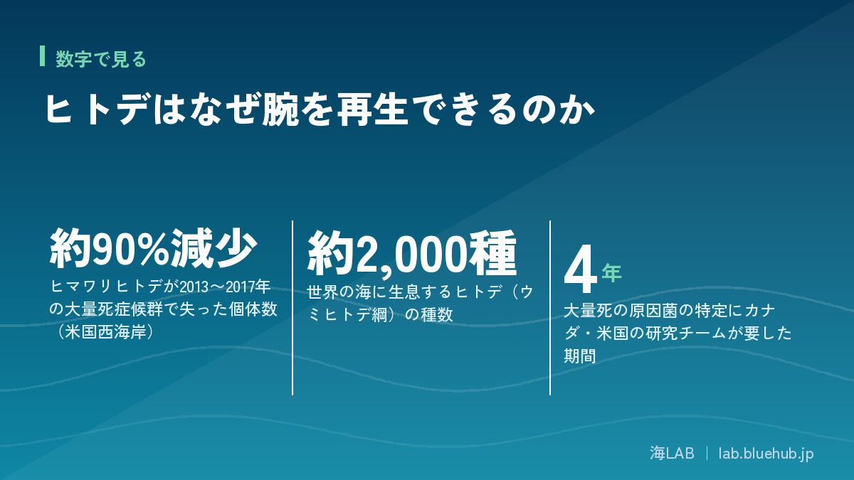
Regeneration speed varies widely by species, extent of damage, and water temperature. A minor injury near an arm tip may show visible regrowth within a few weeks, while a cut near the arm base, or damage to the disc itself, can take several months to over a year before function is fully restored.
The effect of water temperature and season
Like many marine invertebrates, sea star regeneration depends on metabolic rate, so it tends to proceed faster in warmer periods - though extreme heat can instead stress the tissue and hinder regeneration. Experiments that surgically amputated sea star arms under captive conditions have also reported that water temperature and nutritional conditions affect how regeneration proceeds.
The breeding season is another factor worth noting. Many sea stars spawn and release sperm at a fixed time of year, channeling much of their energy into reproduction. When the breeding season overlaps with a regeneration period, the animal faces a kind of trade-off in how it allocates limited internal energy between "reproduction" and "repair," and captive observations suggest that regeneration tends to proceed somewhat more slowly in individuals that are also breeding.
The road to being "fully back to normal"
Even when an arm's shape appears to have recovered within a short time on the outside, internal functions - tube-foot movement, plate density, gonad development - take considerably longer to fully recover than outward appearance does. Researchers in this field stress the importance of not conflating visible regrowth with functional regeneration.
- Early regeneration (days to weeks): the wound is covered by coelomocytes, bleeding stops, and a regeneration bud forms
- Mid regeneration (weeks to months): the bud differentiates into muscle, skeletal plates, skin, and other tissue, and the arm's outer shape gradually takes form
- Late regeneration (months to over a year): coordinated tube-foot movement, pigmentation, and gonad function fully recover until the arm is indistinguishable from the original
Regeneration, then, is not one continuous straight line but a process in which different cells and mechanisms take the lead at each stage. Speed also varies with an individual's age and nutritional condition, and a "regenerating sea star" spotted in the wild is really just a snapshot of one point along this long process.
The research frontier: nerve cord regeneration and a newly found cell population
Sea stars have no brain, but the "radial nerve cords" that extend outward from the ring-shaped "nerve ring" function as the nerve trunks that control arm movement. A study published in an academic journal in 2023 tracked, in detail, the relationship between nerve regeneration and the recovery of motor function after surgically severing this radial nerve cord.
Movement returns as the nerve recovers
The study used a European sea star, Marthasterias glacialis, tracking anatomical and behavioral changes at fine time points after severing the radial nerve cord - one hour, and one, two, eight, and fourteen days later. The team confirmed that coordinated tube-foot movement gradually recovered as the radial nerve cord regenerated, and found that a previously unreported type of coelomocyte population - cells involved in immunity and repair - appeared in the later stage of regeneration. This shows that sea stars do not simply proliferate cells but mobilize different cell populations at different phases of regeneration.
The same study also used mass spectrometry to reveal that the expression levels of 528 different proteins changed significantly in the regenerating nerve tissue, including, for the first time in a regenerating echinoderm nervous system, functional protein groups known to be involved in axon regeneration in other animal species. Behind what looks like the simple return of movement, a great many genes and proteins are working in concert at the molecular level.
Another finding from nerve-regeneration research
A research team at Carnegie Mellon University has also reported that when the neural tissue corresponding to a sea star's "brain" is injured, surrounding cells re-express Sox2, a gene active early in development, restarting the neurogenesis program to build new neurons. Because vertebrates largely lose the ability to regenerate neurons once they reach adulthood, this capacity in sea stars continues to draw the interest of regeneration researchers.
Why sea stars make a good regeneration model
- Their simple body plan makes it easy to observe damage and regeneration at the level of a single "module," the arm
- They show processes shared with vertebrate regeneration, including dedifferentiation, stem-cell activation, and immune response
- They are relatively easy to keep and observe, and regenerating individuals are often findable in wild populations too
Regenerative power seen even at the larval stage
Sea star regeneration is not limited to adults. In studies that surgically cut planktonic sea star larvae, both the front and back fragments were shown to independently go through wound healing, tissue rearrangement, and reconstruction of body-axis patterning, each continuing to swim as an independent larva. The research team showed that some of the same genes used early in development to set the body's axes (front-back, dorsal-ventral) are reused during regeneration - an example of what is thought to be a strategy common across many animals: repurposing the developmental program for regeneration.
What made this research particularly notable was that the same team compared the larval whole-body regeneration process to regeneration processes they had separately studied in other, distantly related animals such as arthropods and annelids. Even across animals separated widely on the tree of life, the basic steps of healing a wound, rebuilding tissue, and reconstructing body axes show many similarities, suggesting that the basic "design" for rebuilding a whole body may be an ancient strategy broadly conserved across the animal kingdom.
Is it true that "cutting a sea star makes more sea stars"?
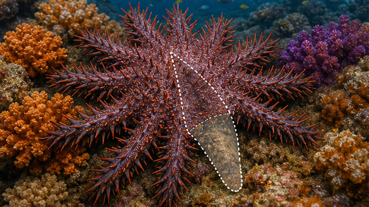
Many people have heard the claim that "sea stars don't die when you cut them - cut one and you get more." This popular belief is half right and half wrong. As described above, a fragment that includes part of the disc can potentially grow into a new individual, but a fragment without disc tissue, or one whose disc is badly damaged, cannot regenerate and will die. In other words, cutting one does not automatically produce more.
Why culling can backfire
A prime example of this misunderstanding causing real harm is the response to crown-of-thorns starfish outbreaks, which devastate coral reefs. When divers try to control crown-of-thorns starfish by chopping them up underwater, fragments that include part of the disc can survive and regenerate, and coral-reef conservation efforts in various regions have reported cases where culling efforts inadvertently increased the population instead. That injecting acetic or formic acid to kill the whole animal has become the mainstream culling method today reflects an understanding of exactly this regeneration risk.
Methods once tried included hauling the animals ashore for disposal, or cutting and sinking them on the spot, but reports of cut fragments regenerating piled up, and crown-of-thorns spines are venomous, posing an injury risk to workers as well. As a result, the internationally standard method today is for divers to inject each individual with a chemical one at a time. This history itself is often cited in coral-reef conservation circles as a cautionary tale of a countermeasure backfiring because regenerative ability was not properly understood.
Not "multiplying" but "occasionally surviving"
To put it scientifically, a sea star is not an animal that "multiplies when cut" but one where "only fragments meeting the right conditions occasionally survive and regenerate." Understanding this distinction matters for avoiding unnecessary population increases during culling or fishery operations.
Lessons for fisheries and conservation work
- Physically chopping an animal up as a culling method risks leaving disc fragments behind that can regenerate and multiply
- Injecting chemicals such as acetic or formic acid to kill the whole individual is now the recommended approach
- For culling other regenerative pest species too, it's important to understand the target's regenerative capacity before choosing a method
The acid-injection method involves a diver injecting each individual with chemicals that break down the animal's own tissue, ensuring it dies completely. It takes more effort, but there is no risk of surviving fragments regenerating, and the impact on surrounding coral and other organisms is relatively small - which is why it has been adopted by coral-reef conservation programs worldwide, including on Australia's Great Barrier Reef. The image of sea stars as creatures that "won't die no matter how you cut them" tends to dominate, but choosing a method grounded in accurate understanding makes culling work that reliably delivers results.
Regenerative power is no guarantee: the wasting-disease pandemic
High regenerative ability at the individual level does not guarantee a species' survival. The textbook example is the mass die-off that struck the large sunflower sea star (Pycnopodia helianthoides) of the North American Pacific coast.
A mysterious mass die-off since 2013
Starting in 2013, along the North American west coast from California to Alaska, sea stars developed ulcer-like lesions on their skin, lost arms that seemed to tear off, and wasted away as if melting - a large-scale event now known as Sea Star Wasting Disease. According to NOAA Fisheries, the sunflower sea star population fell by roughly 80-90% because of the disease, and in some areas across its broad range it disappeared almost entirely. In 2021 the International Union for Conservation of Nature (IUCN) classified the species as Critically Endangered.
Ironically, the disease's symptoms superficially resemble the everyday regeneration process a healthy sea star performs. Small lesions and white patches appear on the skin, and tissue begins to break down from there - the very scene where coelomocytes would normally gather to begin repair. But in this disease, tissue breakdown instead proceeds unchecked: within days an arm twists and drops off, and ultimately the whole individual dies, seeming to dissolve. That an animal group known for exceptional regenerative power suffered mass mortality precisely because its own repair and regeneration processes broke down is what drew researchers' attention.
| Stage | Main symptoms |
|---|---|
| Early infection (before onset to a few days) | Activity slows and tube-foot grip weakens; faint white spots or lesions begin appearing on the skin |
| Progression (a few days to about a week) | Arms twist and swell, skin ulcers spread, and parts of the tissue begin to break down |
| Terminal stage (about one to two weeks after onset) | Arms detach on their own and the whole body loses its shape, collapsing until death |
The pathogen identified in 2025
The cause went unidentified for a long time, but a joint research team from the University of British Columbia (UBC), the Hakai Institute, and the University of Washington spent roughly four years investigating and, in August 2025, pinpointed the cause as a new strain (FHCF-3) of the bacterium Vibrio pectenicida. The team isolated and cultured the bacterium from sea star body fluid and confirmed causation by reproducing the symptoms after injecting it into healthy individuals.
Those four years were not an easy road. Given how the symptoms spread like an infectious disease, many researchers initially suspected a virus, and research narrowing down candidates focused on viruses for a time. But without a decisive breakthrough, years passed, until the team shifted its focus and carefully re-examined the levels of bacteria present in sea star coelomic fluid (the body fluid that functions like blood) - finally arriving at the true cause. That multiple research institutions were involved and many candidates were ruled out before the cause was found illustrates just how time-consuming identifying the source of a wildlife disease can be.
Regenerative power doesn't stop a species from going extinct
- An individual's strong arm-level regenerative power and a population-wide collapse from disease are separate problems
- The sunflower sea star has real regenerative ability, yet it was powerless against this pathogen
- Rising water temperatures from climate change may also have helped the pathogen proliferate
Ripple effects across the ecosystem
The sunflower sea star, which can grow to a meter across, was one of the ocean's leading predators of thin-shelled sea urchins. With this predator nearly gone, purple sea urchins and others freed from predation pressure exploded in number, and multiple surveys have reported that kelp forests across a wide stretch of the North American west coast were devoured, leaving barren rock with little life - so-called "urchin barrens." The die-off of a single sea star species became the trigger for a broader ecosystem shift affecting fish, seabirds, and even coastal fisheries, which is why sea star regeneration and reproduction research matters well beyond this one species.
Species other than the sunflower sea star did not escape unharmed either. This wasting disease has been confirmed in at least 20 sea star species along the North American Pacific coast from Alaska to Mexico, and Pisaster ochraceus, known as a keystone species in intertidal ecosystems, was also hit hard. Differences in symptom severity and recovery capacity across species are becoming clearer, and now that the pathogen has been identified, further research into species-by-species susceptibility is expected.
The front line of conservation: captive breeding and the push to return sea stars to the wild
The sunflower sea star is also an important predator of urchins that graze on kelp and other seaweeds. In areas where the sunflower sea star has plummeted, urchins have overpopulated and devoured kelp forests, causing "urchin barrens", a phenomenon reported in multiple regions, positioning this species' conservation as an ecosystem-wide issue.
Captive breeding by a nonprofit research lab
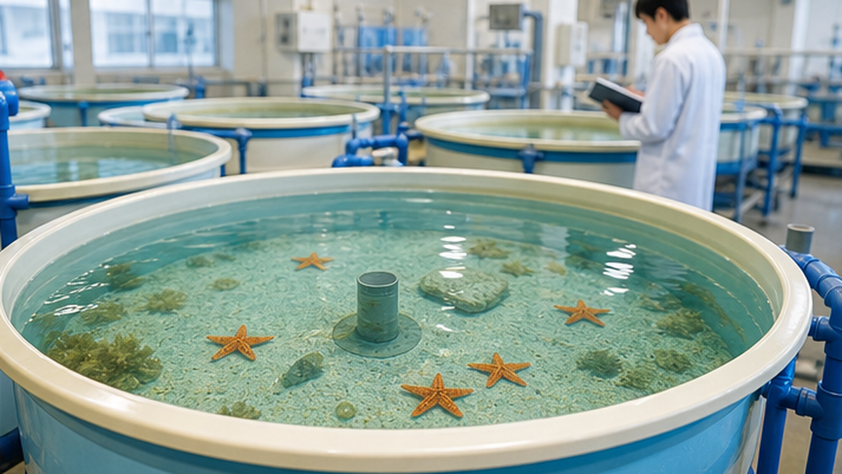
Sunflower Star Laboratory, a nonprofit research organization based in Monterey, California, is working with partners such as the California Academy of Sciences and The Nature Conservancy to captive-breed sunflower sea stars using sperm and eggs collected from adults in human care. According to the organization, a cohort of juveniles raised from gametes collected at Birch Aquarium and elsewhere in 2024 continues to be raised at their facility.
The breeding process uses not only fresh sperm but also frozen, cryopreserved sperm. This helps maintain genetic diversity by drawing on multiple individuals held at different locations, avoiding the risk of ending up with a genetically uniform population by relying only on a small number of surviving animals. Now that the pathogen has been identified, the next challenge is shifting to a more practical stage: how to raise populations that can withstand the pathogen, and how to safely return them to the sea.
The adult broodstock used for breeding is kept spread across multiple aquariums, including Birch Aquarium and the Aquarium of the Pacific, with gametes collected from them brought together at the Monterey facility for fertilization and juvenile rearing. By not relying on a single facility for all broodstock, the program aims to spread the risk so that a disaster or infection at one site doesn't derail the whole effort. Newly hatched juveniles are only a few millimeters across, and it takes many years to reach adult size, so the visible results of this conservation work still lie some way in the future.
The challenge of returning animals to the wild
Even with successful captive breeding, as long as the pathogen remains present in the ocean, releasing captive-raised animals back into the wild carries the risk of reinfection. Research teams are considering selecting for pathogen-resistant individuals and planning staged reintroductions into lower-risk waters, and conservation is shifting its focus from simply "breeding more" to "returning them safely."
Moves toward federal protection in the US
NOAA Fisheries has proposed listing the sunflower sea star as "threatened" under the US Endangered Species Act. If the listing goes through, it would enable legally backed protection measures, including habitat conservation and bycatch prevention. Now that it's clear that individual-level regenerative power alone cannot save a species, there is growing emphasis on combining captive breeding - a technology for increasing individual numbers - with legal protections for capture and habitat.
What sea star research could offer regenerative medicine
Sea star regeneration research is more than an interesting aquarium topic; it also carries real value as basic biology. The process by which dedifferentiated cells redetermine their fate and build the right tissue in the right place shares common ground with basic research into human wound healing and regenerative medicine.
Parallels and differences with newts and planarians
Newts, which regenerate limbs, planarians, which can regenerate even after being cut into dozens of pieces, and sea stars are often mentioned together as the animal kingdom's "star pupils" of regeneration. All three share the use of dedifferentiation and stem-cell-like cells, but sea stars are distinctive in that immune-related coelomocytes are deeply involved in the repair process, tightly linking defense against infection and foreign material with tissue regeneration.
| Animal | What it can regenerate | Main mechanism | Taxonomic group |
|---|---|---|---|
| Sea star | Arms, and under the right conditions a whole individual | Immune response via coelomocytes plus dedifferentiation of muscle and epidermal cells | Echinoderm (invertebrate) |
| Newt | Limbs, tail, eye lens, and more | Dedifferentiation of wound-site cells and formation of a regeneration bud | Vertebrate (amphibian) |
| Planarian | Almost any part of the body (a whole individual from a cut fragment) | Pluripotent stem cells called neoblasts distributed throughout the body | Flatworm (invertebrate) |
Human application is still far off, but the significance is real
Sea star regeneration mechanisms are nowhere near being applied directly in human medicine. But for the fundamental question of why most animals cannot regenerate complex organs while a few can, research on simply organized animals like sea stars keeps offering clues at the genetic level.
Where the research goes next
Now that the pathogen behind the wasting-disease pandemic has been identified, research is shifting toward a deeper question: why can a sea star with such strong regenerative power fail to repair itself against this particular disease and instead collapse? Comparing the immune and regenerative responses of healthy individuals with those of diseased ones at the molecular level could reveal exactly where coelomocyte function breaks down, potentially opening a path toward treatments or breeding for resistant individuals. Sea star regeneration research is both basic science probing the mystery of animal regeneration and applied research directly relevant to a conservation crisis unfolding right now.
The next time you spot a sea star in a tide pool with one arm shorter than the rest, it may help to remember: that's not an incomplete creature, but a snapshot of one living through the process of repairing a wound. Combining a stubborn, individual-level power to regenerate with a species-level vulnerability to disease and environmental change, the sea star reflects, in miniature, both the resilience and the fragility of ocean life.
References and sources
- NOAA Fisheries - Sunflower Sea Star - Official information on the species' biology and population decline
- NOAA Ocean Service - Are starfish really fish? - Explainer on sea star classification and species count
- UBC News - Disease detectives discover cause of sea star wasting disease - August 2025 announcement identifying the pathogen behind the mass die-off
- PMC - Regeneration of starfish radial nerve cord restores animal mobility and unveils a new coelomocyte population - 2023 paper on nerve-cord regeneration and a new coelomocyte population
- PMC - Analysis of sea star larval regeneration reveals conserved processes of whole-body regeneration across the metazoa - Research on whole-body regeneration in larvae
- Seto Marine Biological Laboratory, Kyoto University - "The Regenerative Power of Sea Stars" (PDF) - General-audience explainer on arm regeneration
- Sunflower Star Laboratory - Nonprofit research organization working on captive breeding and conservation of the sunflower sea star
- Puget Sound Institute - Sunflower sea stars certified as 'critically endangered' - Reporting on the IUCN Red List listing
- Carnegie Mellon University - Research Reveals Ways Neurons are Regenerated in Starfish - 2022 research announcement on sea star neuron regeneration
- Hokkaido University Graduate School of Life Science - "Five- and six-armed brittle stars 'bulge' differently" - Research on brittle star arm count and movement coordination
* Listed roughly in order of reliability: government and academic institutions > peer-reviewed papers > specialist organizations > trusted media